CT Scan, Computed Tomography, CT Scan procedure, CT Scan results, CT scan benefits, CT scan Risks, CT Scan limiation
Abdominal CT Scan- Procedure, Risks and Result
Computed tomography (CT scan or CAT scan) is a noninvasive diagnostic imaging procedure that uses a combination of X-rays and computer technology to show cross-sectional images of a specific area of the body.
What is CT Scanning of the Abdomen?
Computed tomography, more commonly known as a CT or CAT scan, is a diagnostic medical imaging test. Like traditional x-rays, it produces multiple images or pictures of the inside of the body.
A CT scan generates images that can be reformatted in multiple planes. It can even generate three-dimensional images. Your doctor can review these images on a computer monitor, print them on film or via a 3D printer, or transfer them to a CD or DVD.
CT images of internal organs, bones, soft tissue, and blood vessels provide greater detail than traditional x-rays. This is especially true for soft tissues and blood vessels.
Common uses of CT Scan
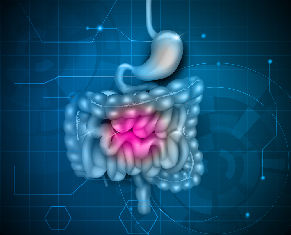
An abdominal CT scan makes detailed pictures of the structures inside your belly very quickly.
This test may be used to look for:
- Cause of blood in the urine
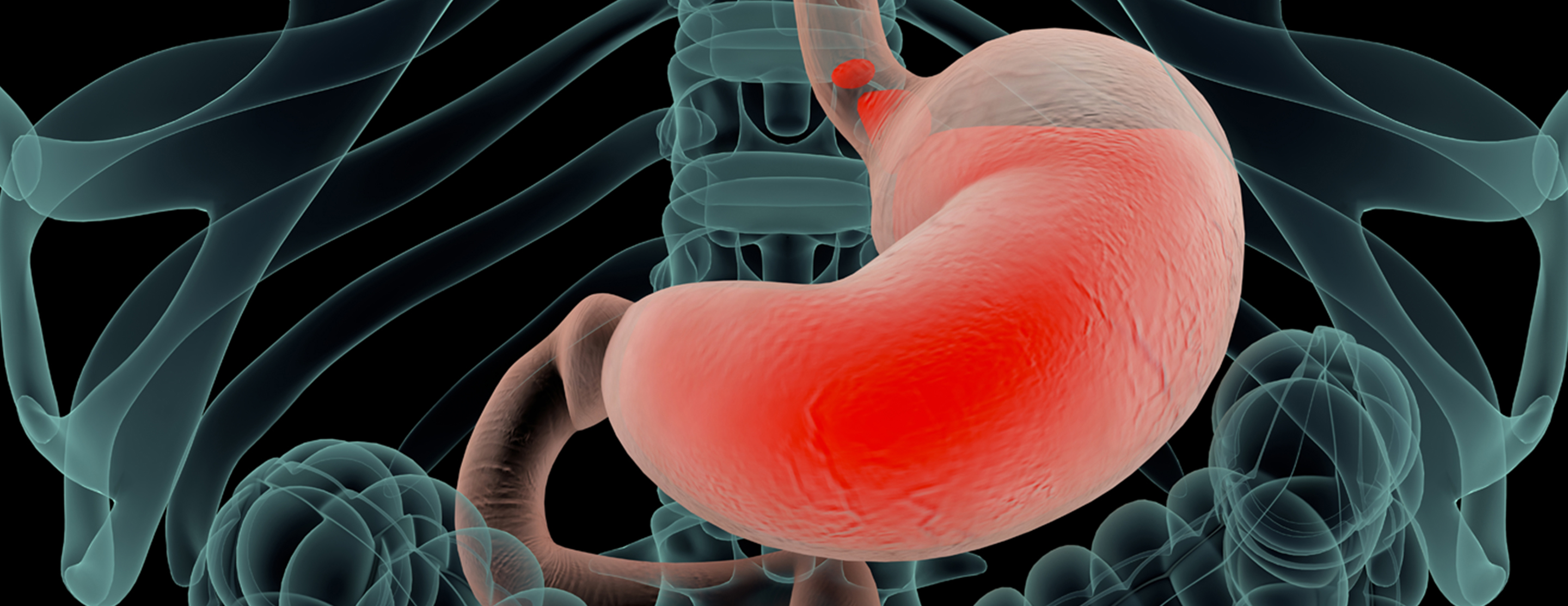
- Cause of abdominal pain or swelling
- Cause of abnormal blood test results such as liver or kidney problems
- Hernia
- Cause of a fever
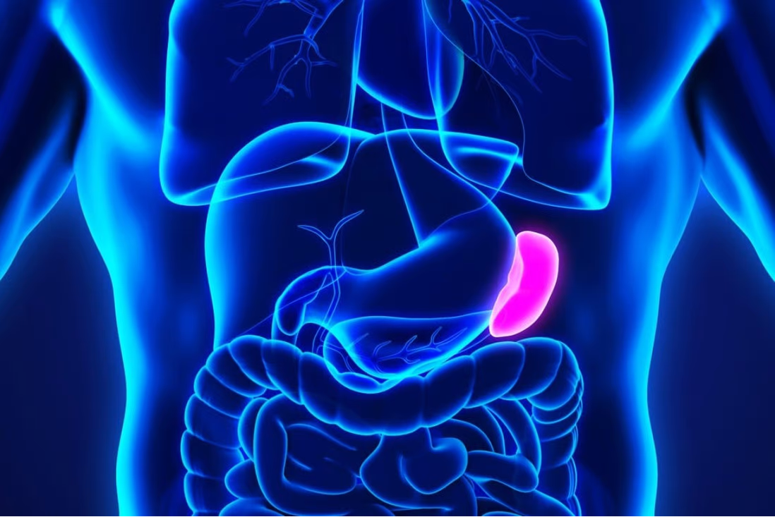
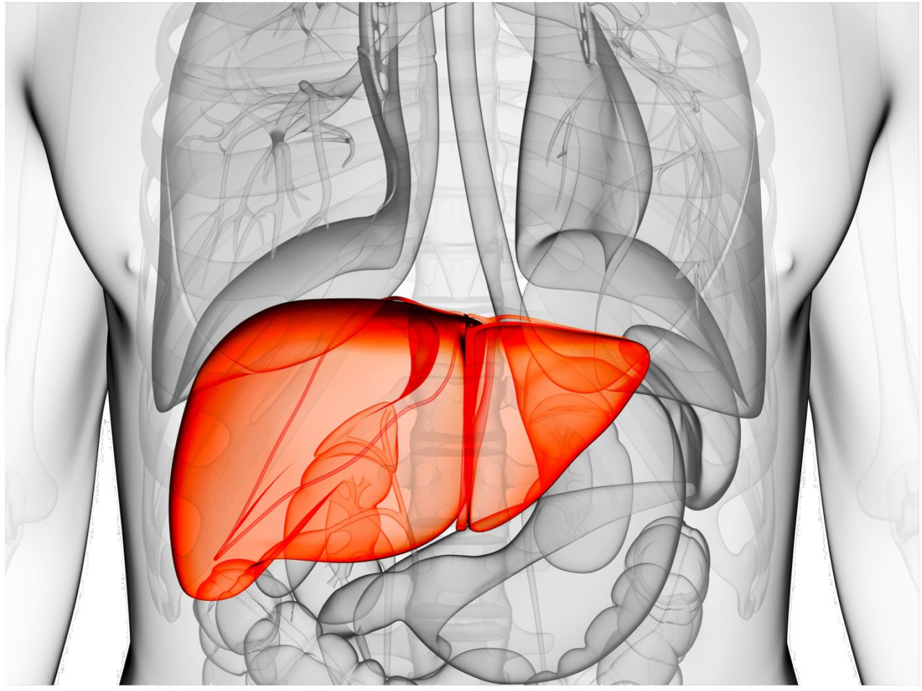
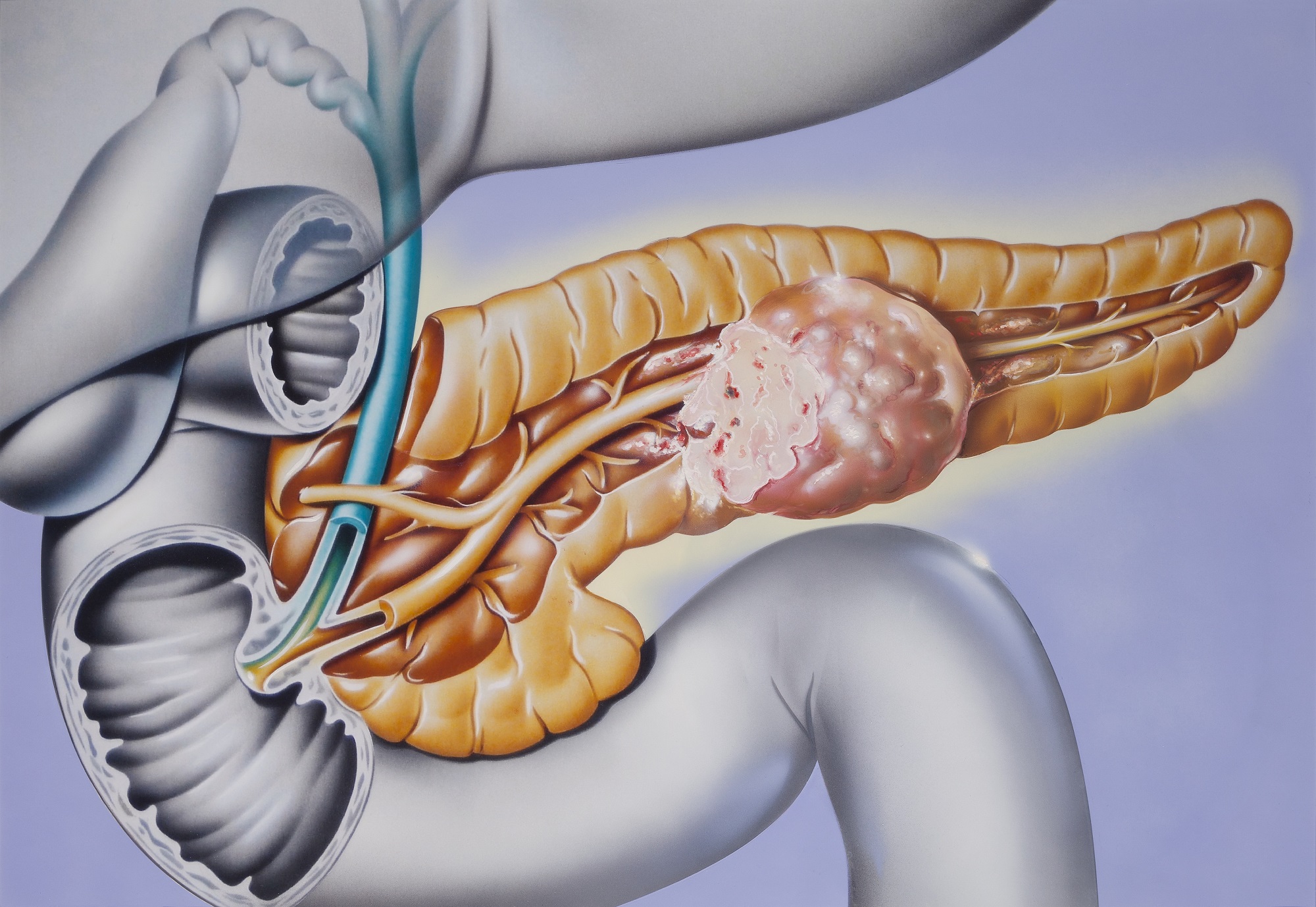
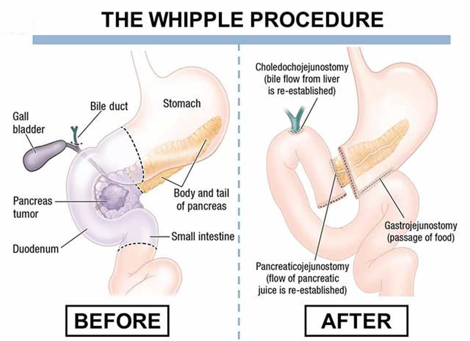
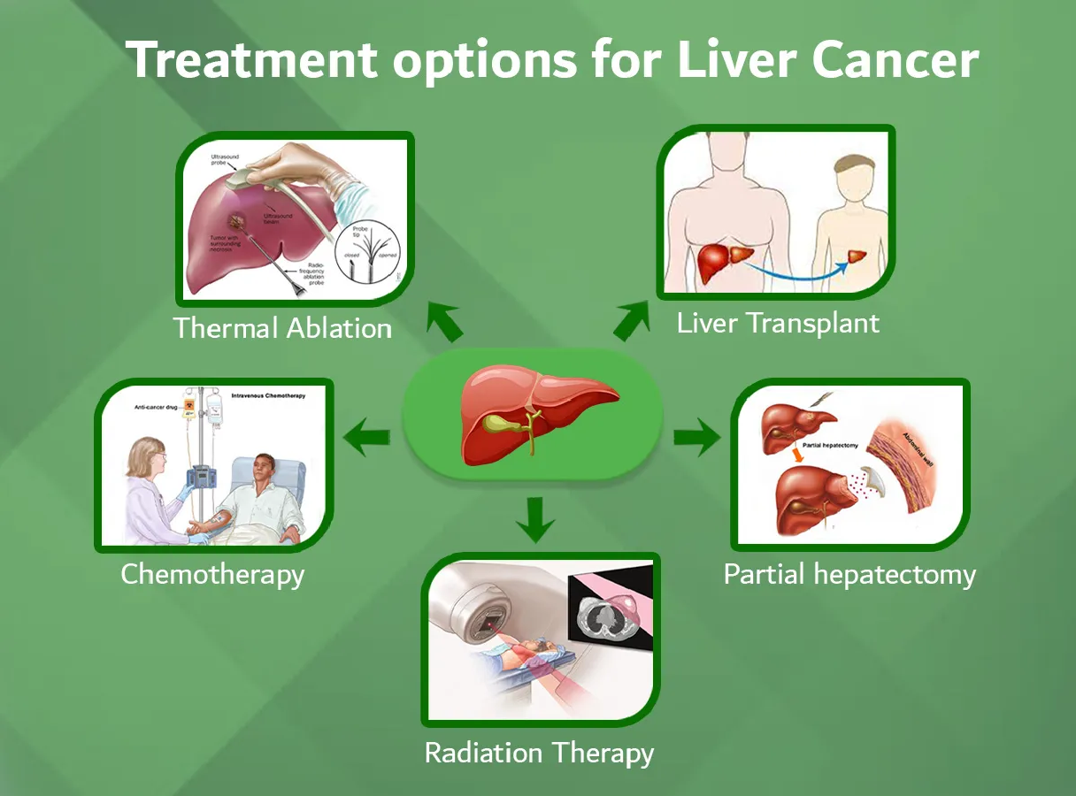
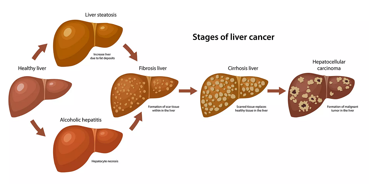
- Masses and tumors, including cancer of liver, kidneys, pancreas, ovaries and bladder as well as lymphoma
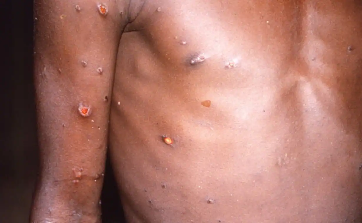
- Infections or injury
- Kidney stones
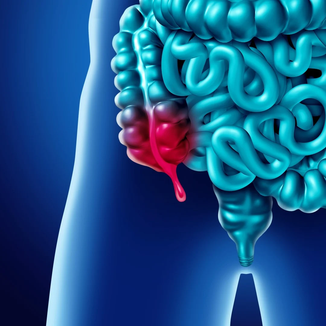
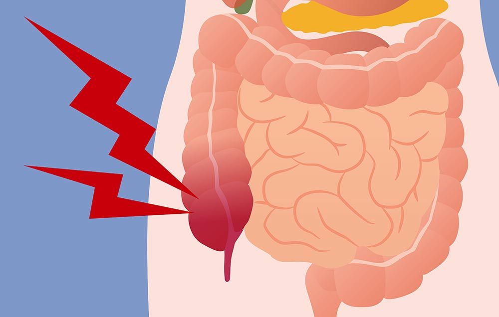
- Appendicitis
Doctors also use CT scanning of the abdomen to:
- Guide biopsies and other procedures such as abscess drainages and minimally invasive tumor treatments.
- Plan for and assess the results of surgery, such as organ transplants.
- Stage, plan and properly administer radiation treatments for tumors as well as monitor response to chemotherapy.
CT scan vs. MRI vs. X-ray
You may have heard of other imaging exams and wonder why your doctor chose a CT scan over other options.
Your doctor may choose a CT scan over an MRI (magnetic resonance imaging) scan because a CT scan is faster than an MRI. Plus, if you’re uncomfortable in small spaces, a CT scan would likely be a better choice.
An MRI requires you to be inside an enclosed space while loud noises occur all around you. In addition, an MRI is more expensive than a CT scan.
Your doctor may choose a CT scan over an X-ray because it provides more detail than an X-ray does. A CT scanner moves around your body and takes pictures from many different angles. An X-ray takes pictures from one angle only.
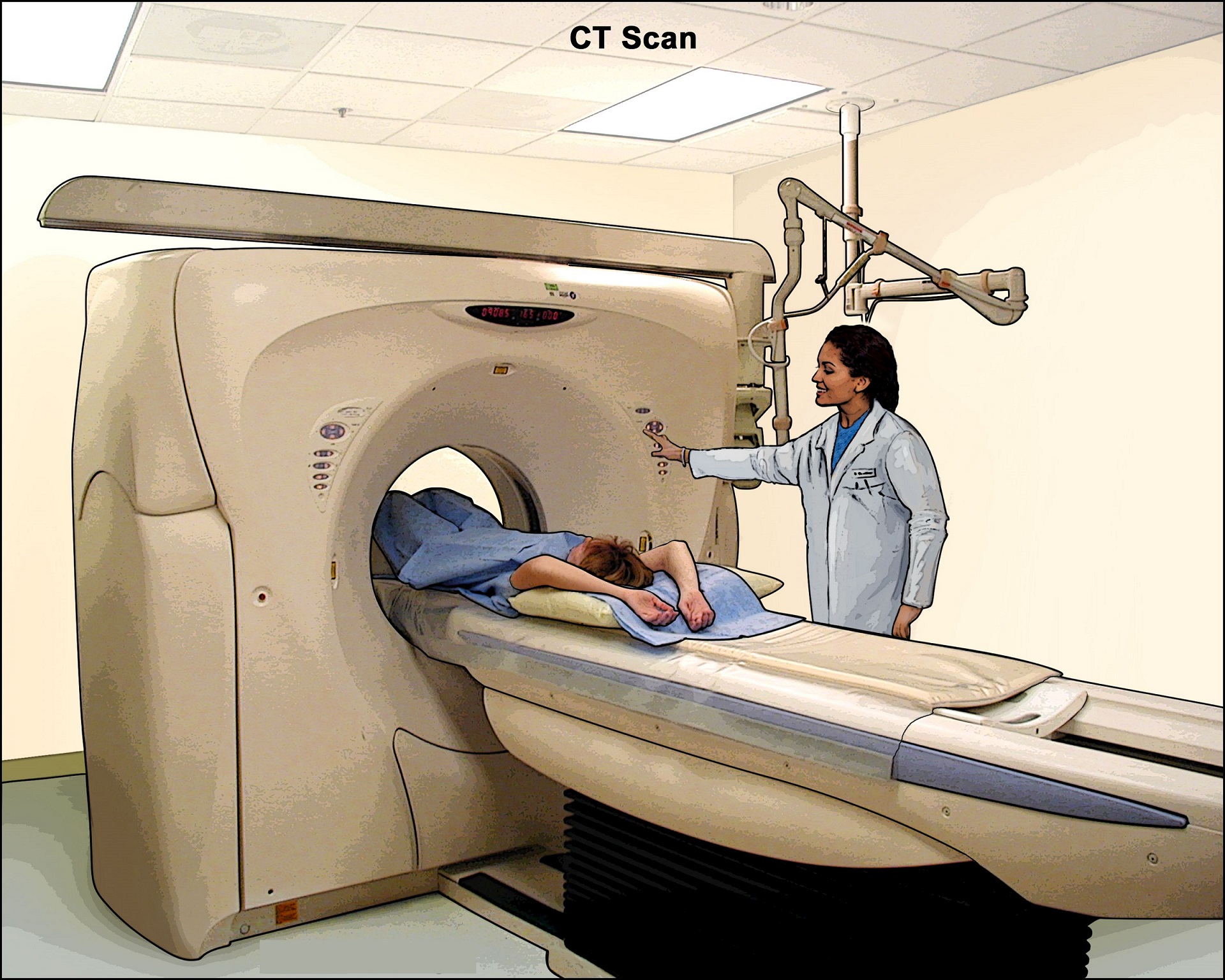
What does the CT equipment look like?
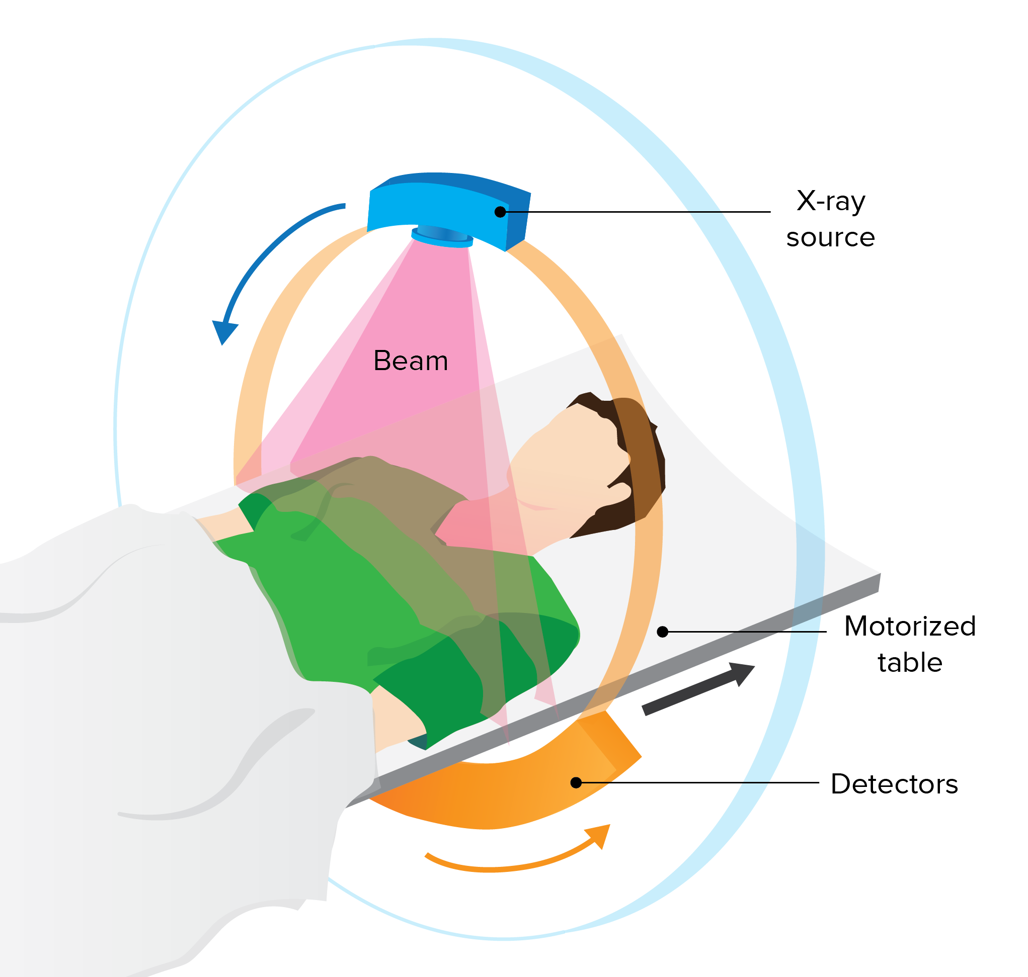
The CT scanner is typically a large, donut-shaped machine with a short tunnel in the center. You will lie on a narrow table that slides in and out of this short tunnel. Rotating around you, the x-ray tube and electronic x-ray detectors are located opposite each other in a ring, called a gantry. The computer workstation that processes the imaging information is in a separate control room. This is where the technologist operates the scanner and monitors your exam in direct visual contact. The technologist will be able to hear and talk to you using a speaker and microphone.
How does the procedure work?
In many ways, a CT scan works like other x-ray exams. Different body parts absorb x-rays in different amounts. This difference allows the doctor to distinguish body parts from one another on an x-ray or CT image.
A conventional x-ray exam directs a small amount of radiation through the body part under examination. A special electronic image recording plate captures the image. Bones appear white on the x-ray. Soft tissue, such as the heart or liver, shows up in shades of gray. Air appears black.
With CT scanning, several x-ray beams and electronic x-ray detectors rotate around you. These measure the amount of radiation being absorbed throughout your body. Sometimes, the exam table will move during the scan. A special computer program processes this large volume of data to create two-dimensional cross-sectional images of your body. The system displays the images on a computer monitor. CT imaging is sometimes compared to looking into a loaf of bread by cutting the loaf into thin slices. When the computer software reassembles the image slices, the result is a very detailed multidimensional view of the body's interior.
Nearly all CT scanners can obtain multiple slices in a single rotation. These multi-slice (multidetector) CT scanners obtain thinner slices in less time. This results in more detail.
Modern CT scanners can image large sections of the body in just a few seconds, and even faster in small children. Such speed is beneficial for all patients. Speed is especially beneficial for children, the elderly, and critically ill – anyone who finds it difficult to stay still, even for the brief time necessary to obtain images.
For children, the radiologist will adjust the CT scanner technique to their size and the area of interest to reduce the radiation dose.
Some CT exams use a contrast material to enhance visibility in the body area under examination.
How is the procedure performed?
The technologist begins by positioning you on the CT exam table, usually lying flat on your back. They may use straps and pillows to help you maintain the correct position and remain still during the exam.
Many scanners are fast enough to scan children without sedation. In special cases, children who cannot hold still may need sedation. Motion may cause blurring of the images and degrade image quality the same way that it affects photographs.
The exam may use contrast material, depending on the type of exam. If so, it will be swallowed, injected through an intravenous line (IV) or, rarely, administered by enema.
Next, the table will move quickly through the scanner to determine the correct starting position for the scans. Then, the table will move slowly through the machine for the actual CT scan. Depending on the type of CT scan, the machine may make several passes.
The technologist may ask you to hold your breath during the scanning. Any motion, including breathing and body movements, can lead to artifacts on the images. This loss of image quality can resemble the blurring seen on a photograph taken of a moving object.
When the exam is complete, the technologist will ask you to wait until they verify that the images are of high enough quality for accurate interpretation by the radiologist.
The CT exam usually takes just a few minutes. However, if you need to drink oral contrast, the imaging centre will ask you to arrive approximately two hours prior to your scan time. Or, they may instruct you to start drinking the contrast at home prior to arriving.
How to Prepare for the Test
Let your health care provider know if you have ever had a reaction to contrast. You may need to take medicines before the test to safely receive this substance.
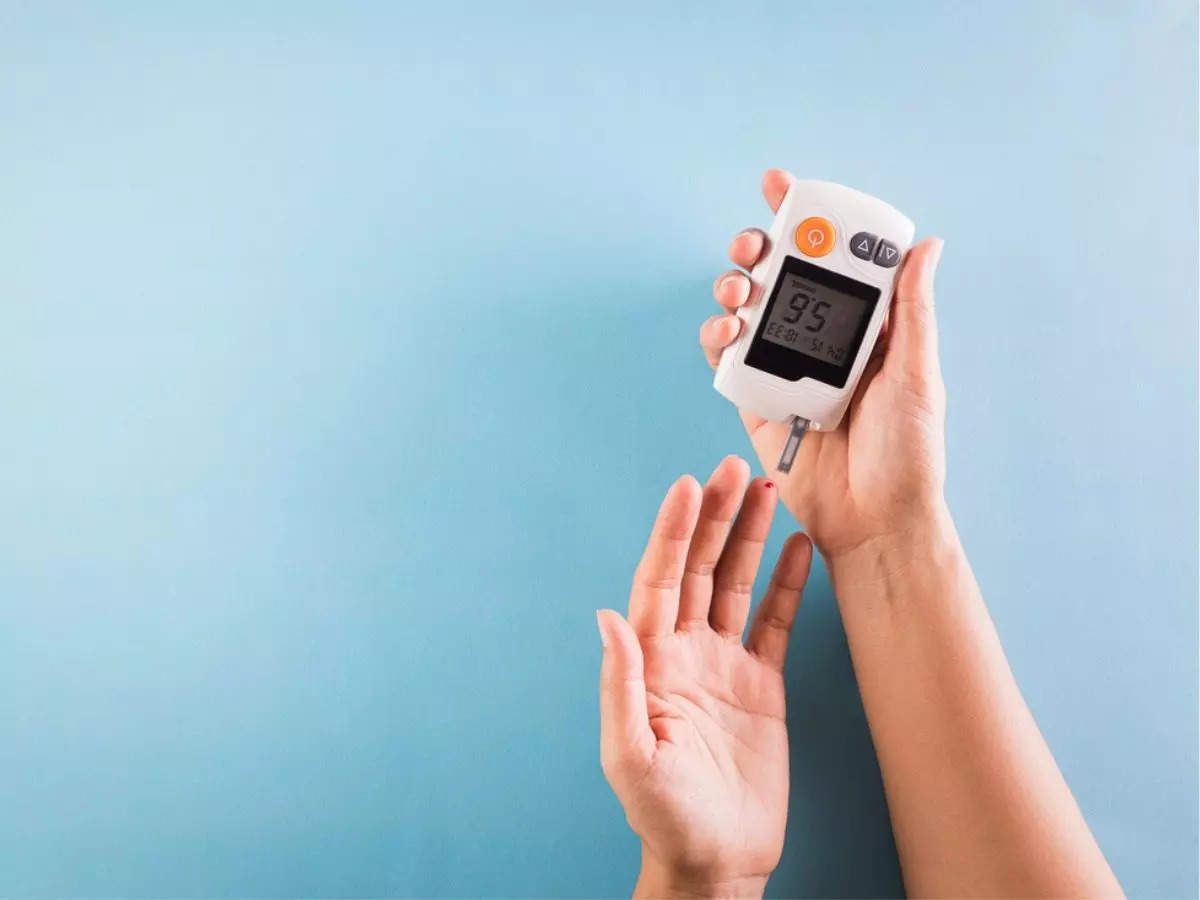
Before receiving the contrast, tell your provider if you take the diabetes medicine metformin. People taking this medicine may have to stop taking it for a while before the test.
Let your provider know if you have any kidney problems. The IV contrast can worsen kidney function.
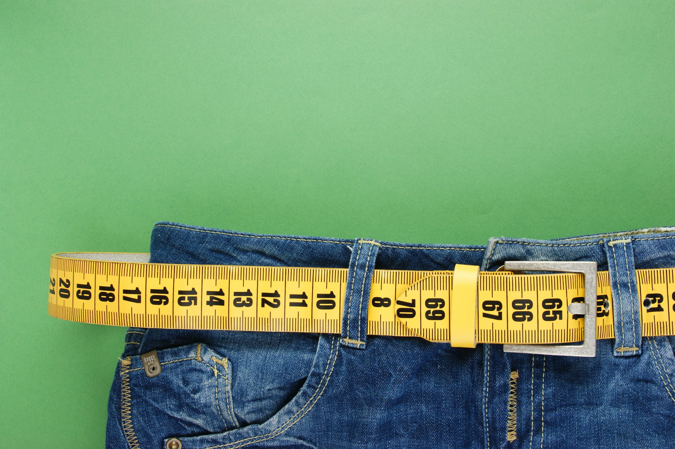
Too much weight can damage the scanner. Find out if the CT machine has a weight limit if you are obese.
You will need to take off your jewelry and wear a hospital gown during the study.
How the Test will Feel
Lying on the hard table may be a little bit uncomfortable.
If you have contrast through a vein (IV), you may have:
- Slight burning sensation
- Metallic taste in the mouth
- Warm flushing of the body
These feelings are normal and go away within a few seconds.
What will I experience during and after the procedure?
CT exams are generally painless, fast, and easy. Multidetector CT reduces the amount of time that the patient needs to lie still.
Though the scan is painless, you may have some discomfort from remaining still for several minutes or from placement of an IV. If you have a hard time staying still, are very nervous, anxious, or in pain, you may find a CT exam stressful. The technologist or nurse, under the direction of a doctor, may offer you some medication to help you tolerate the CT exam.
If the exam uses iodinated contrast material, your doctor will screen you for chronic or acute kidney disease. The doctor may administer contrast material intravenously (by vein), so you will feel a pin prick when the nurse inserts the needle into your vein. You may feel warm or flushed as the contrast is injected. You also may have a metallic taste in your mouth. This will pass. You may feel a need to urinate. However, these are only side effects of the contrast injection, and they subside quickly.
If you swallow oral contrast material, you may find the taste mildly unpleasant. However, most patients can easily tolerate it. If you receive an enema, you can expect to experience a sense of abdominal fullness. You may also feel an increasing need to expel the liquid. If so, be patient; the mild discomfort will not last long.
Many patients also receive an iodine-based contrast material intravenously (injected into a vein) to help evaluate blood vessels and organs such as the liver, kidneys, and pancreas.
When you enter the CT scanner, you may see special light lines projected onto your body. These lines help ensure that you are in the correct position on the exam table. With modern CT scanners, you may hear slight buzzing, clicking and whirring sounds. These occur as the CT scanner's internal parts, not usually visible to you, revolve around you during the imaging process.
You will be alone in the exam room during the CT scan, unless there are special circumstances. For example, sometimes a parent wearing a lead shield may stay in the room with their child. However, the technologist will always be able to see, hear and speak with you through a built-in intercom system.
With pediatric patients, a parent may be allowed in the room but may need to wear a lead apron to minimize radiation exposure.
After a CT exam, the technologist will remove your intravenous line. They will cover the tiny hole made by the needle with a small dressing. You can return to your normal activities immediately.
Who interprets the results and how do I get them?
A radiologist, a doctor specially trained to supervise and interpret radiology exams, will analyze the images. The radiologist will send an official report to the doctor who ordered the exam.
You may need a follow-up exam. If so, your doctor will explain why. Sometimes a follow-up exam further evaluates a potential issue with more views or a special imaging technique. It may also see if there has been any change in an issue over time. Follow-up exams are often the best way to see if treatment is working or if a problem needs attention.
What are the benefits of CT Scan?
- Viewing a CT scan, an experienced radiologist can diagnose many causes of abdominal pain or injury from trauma with very high accuracy. This allows for faster treatment and often eliminates the need for additional, more invasive diagnostic procedures.
- When pain is caused by infection and inflammation, the speed, ease and accuracy of a CT exam can reduce the risk of serious complications. Such complications may include those caused by a burst appendix or an infected fluid collection and the subsequent spread of infection.
- CT scanning is painless, noninvasive, and accurate.
- A major advantage of CT is its ability to image bone, soft tissue, and blood vessels all at the same time.
- Unlike conventional x-rays, CT scanning provides very detailed images of many types of tissue as well as the lungs, bones, and blood vessels.
- CT exams are fast and simple. In emergency cases, they can reveal internal injuries and bleeding quickly enough to help save lives.
- CT has been shown to be a cost-effective imaging tool for a wide range of clinical problems.
- CT is less sensitive to patient movement than MRI.
- Unlike MRI, an implanted medical device of any kind will not prevent you from having a CT scan.
- CT imaging provides real-time imaging, making it a good tool for guiding needle biopsies and needle aspirations. This is particularly true of procedures involving the lungs, abdomen, pelvis, and bones.
- A diagnosis via CT scan may eliminate the need for exploratory surgery and surgical biopsy.
- No radiation remains in a patient's body after a CT exam.
- The x-rays used for CT scanning should have no immediate side effects.
What are the risks of CT Scan?
- There is always a slight chance of cancer from excessive exposure to radiation. However, the benefit of an accurate diagnosis far outweighs the risk involved with CT scanning.
- The radiation dose for this procedure varies. See the Radiation Dose in X-Ray and CT Exams page for more information about radiation dose.
- Women should always tell their doctor and x-ray or CT technologist if there is any chance they are pregnant.
- Doctors do not generally recommend CT scanning for pregnant women unless medically necessary because of potential risk to the unborn baby.
- The risk of serious allergic reaction to contrast materials that contain iodine is extremely rare, and radiology departments are well-equipped to deal with reactions.
- IV contrast manufacturers indicate mothers should not breastfeed their babies for 24-48 hours after contrast material is given.
Because children are more sensitive to radiation, they should have a CT exam only if it is essential for making a diagnosis. They should not have repeated CT exams unless necessary. CT scans in children should always be done with low-dose technique.
Radiology departments tailor the radiation dose for CT scans, especially when scanning children. This helps ensure that the benefits of the scan far outweigh any possible risks from the exposure to diagnostic radiation.
What are the limitations of Abdominal CT?
A person who is very large may not fit into the opening of a conventional CT scanner. Or, they may be over the weight limit. Industry standard table weight limits for CT is usually 450 Ibs (205 kg). Newer larger CT scanners are currently available which can accommodate patients weighing up to 680 Ibs (308.4 kg). These table weight limits exist because the table movement mechanics can be limited by the weight of the patient.
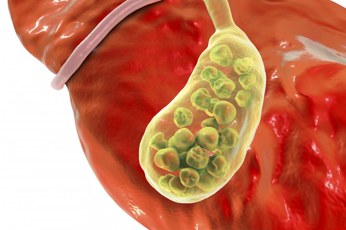
CT scanning of the abdomen may not be as sensitive in identifying gallstones as ultrasound of the abdomen.
Doctors prefer alternate imaging techniques such as plain films, gastrointestinal (GI) contrast exams and ultrasound for evaluating acute abdominal conditions in babies, such as vomiting or blood in stool.
For some conditions, including but not limited to some liver, kidney, pancreatic, uterine, or ovarian abnormalities, evaluation and diagnosis with MRI may be preferable to CT scanning.