ultrasound, ultrasonography, sonography, ultrasound, USG, Ultrasound procedure, ultrasound result
Ultrasound/ Ultrasonography (USG)- Purpose, Procedure, Preparation and more
Ultrasound is a medical imaging technique that uses high-frequency sound waves to produce images of internal body structures.
What is Ultrasound/ Ultrasonography?
Diagnostic ultrasound, also called sonography or diagnostic medical sonography, is an imaging method that uses sound waves to produce images of structures within your body. The images can provide valuable information for diagnosing and directing treatment for a variety of diseases and conditions.
Most ultrasound examinations are done using an ultrasound device outside your body, though some involve placing a small device inside your body.
Why is Ultrasound done?
Ultrasound is used for many reasons, including to:
- View the uterus and ovaries during pregnancy and monitor the developing baby's health
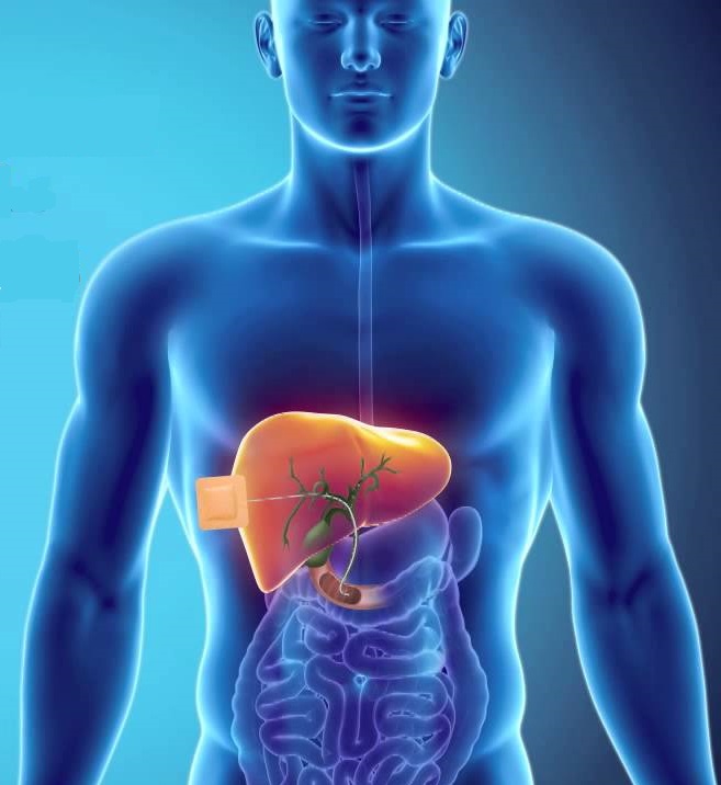
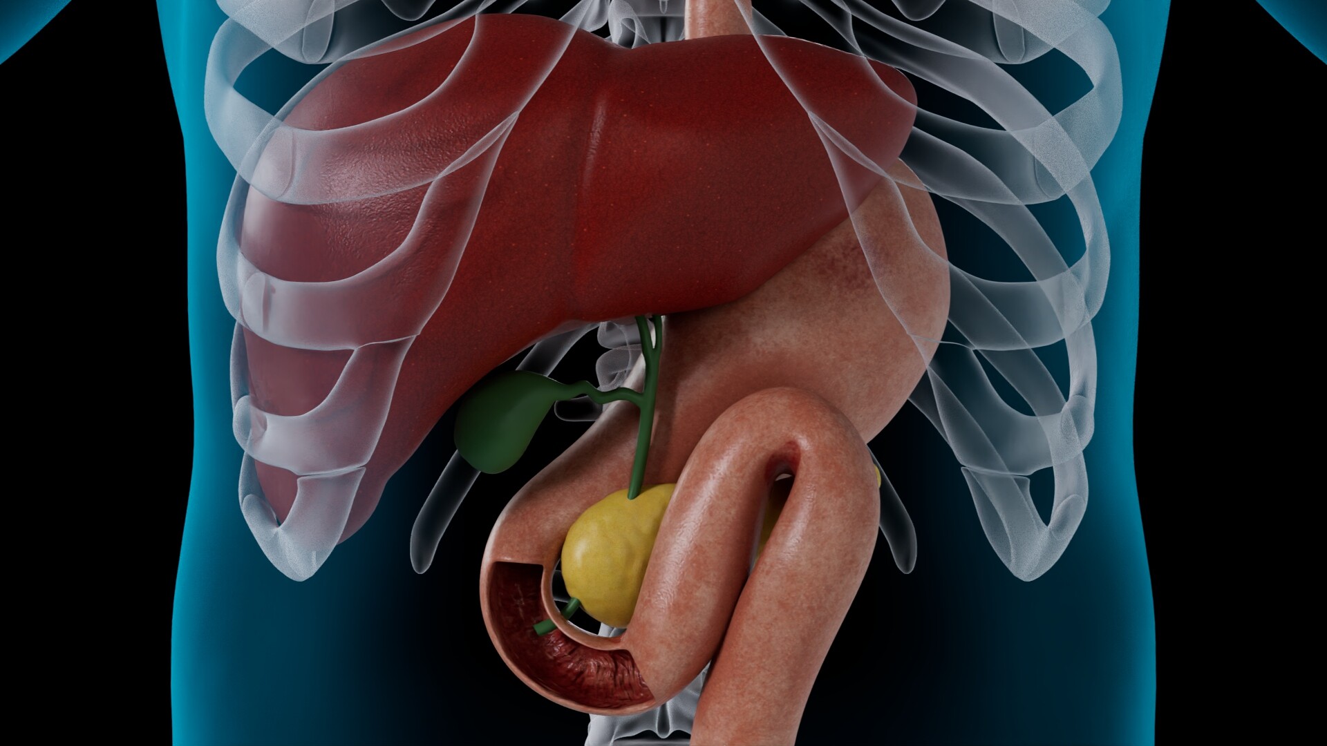
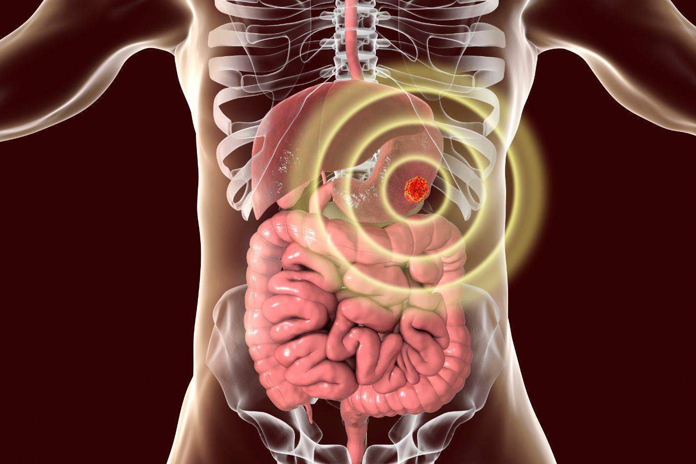
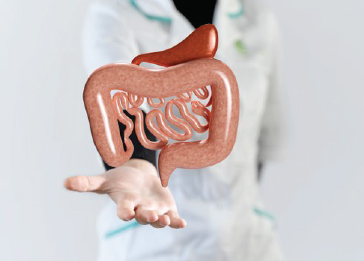
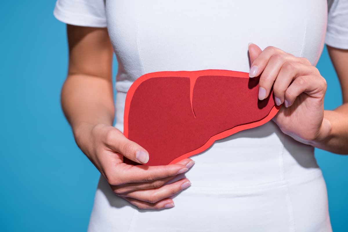
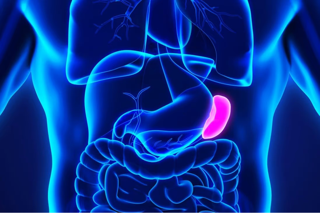
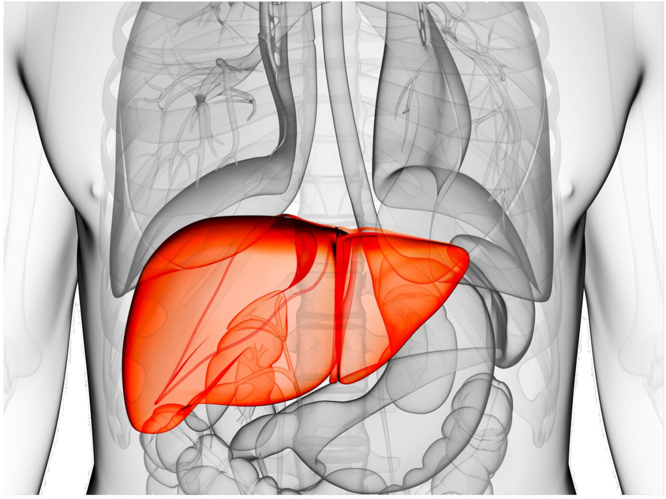
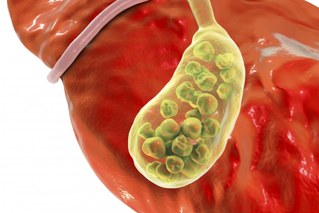
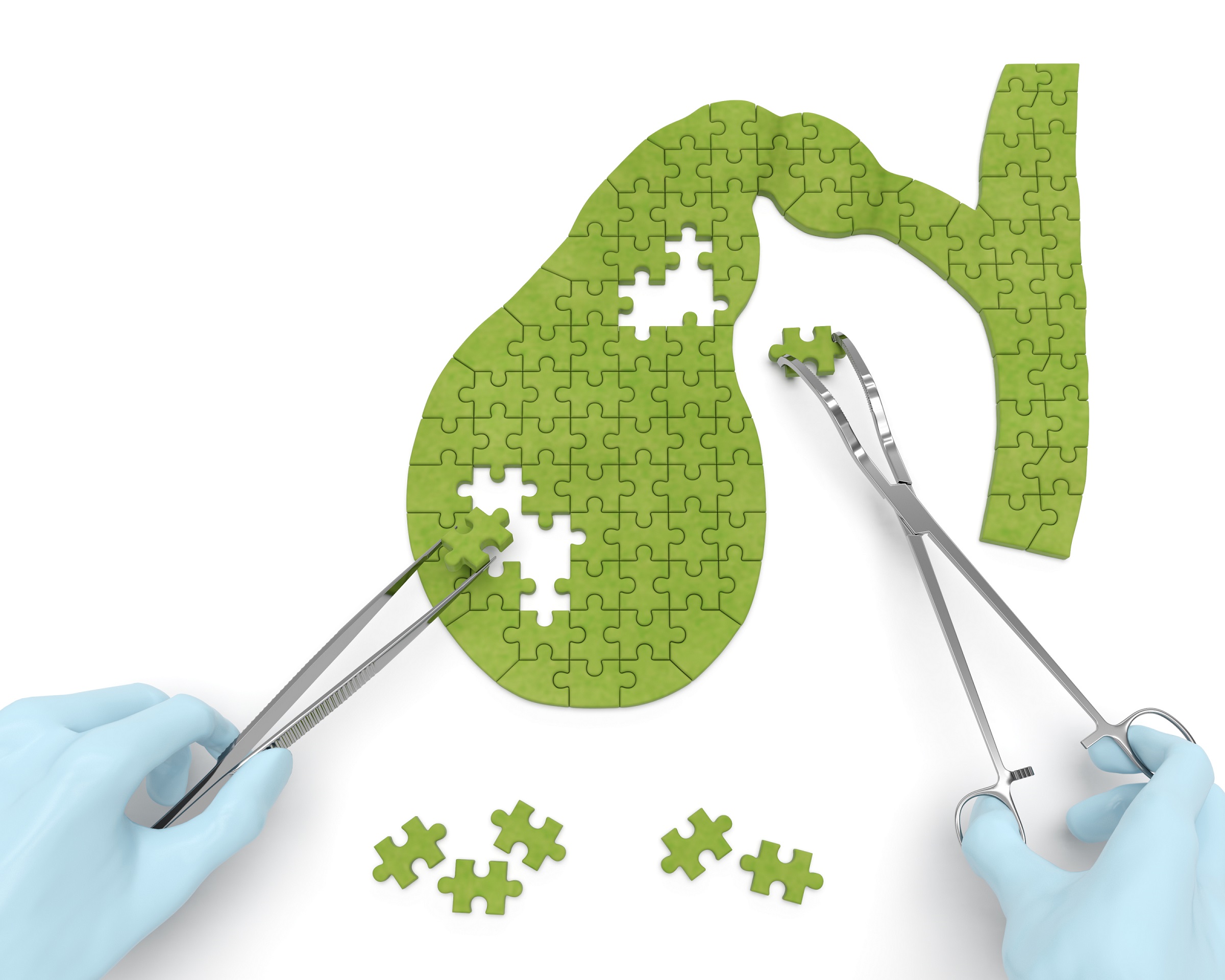
- Diagnose gallbladder disease
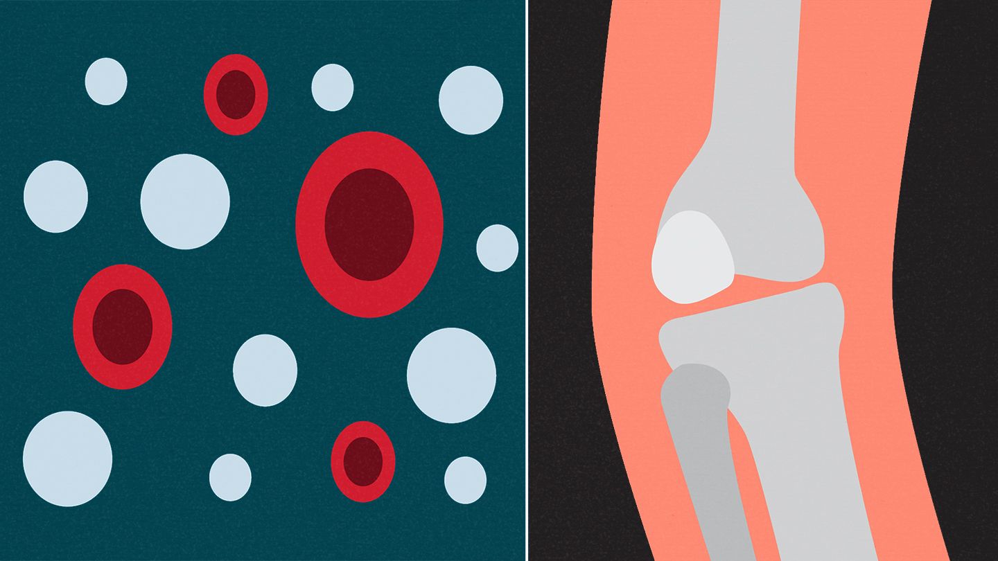
- Evaluate blood flow
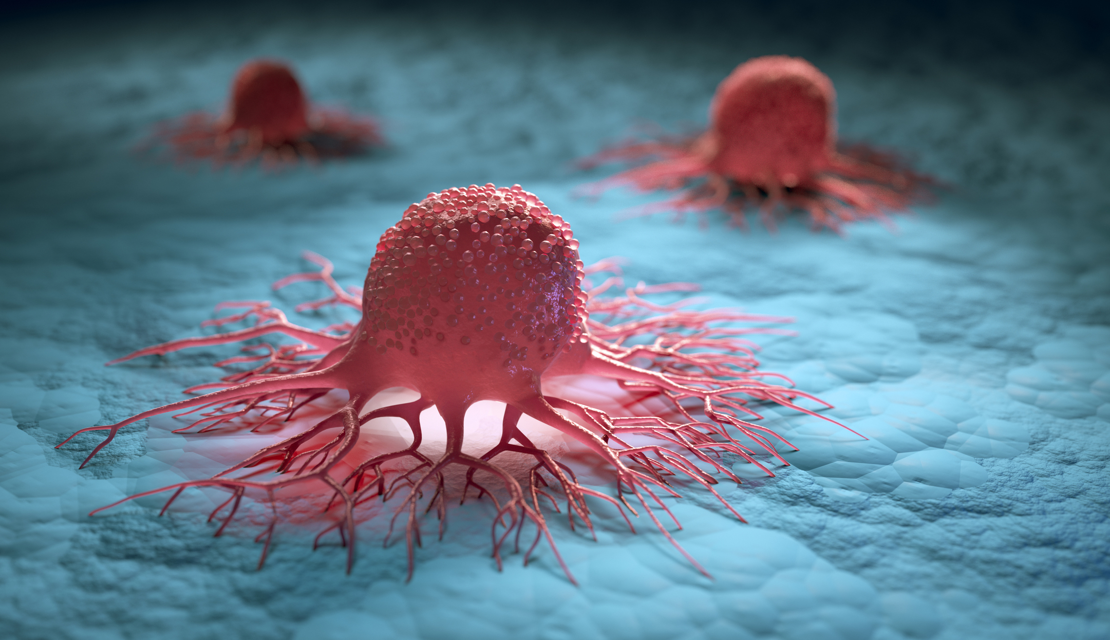
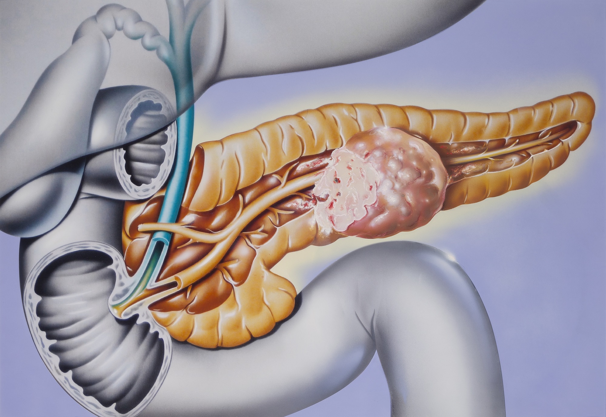
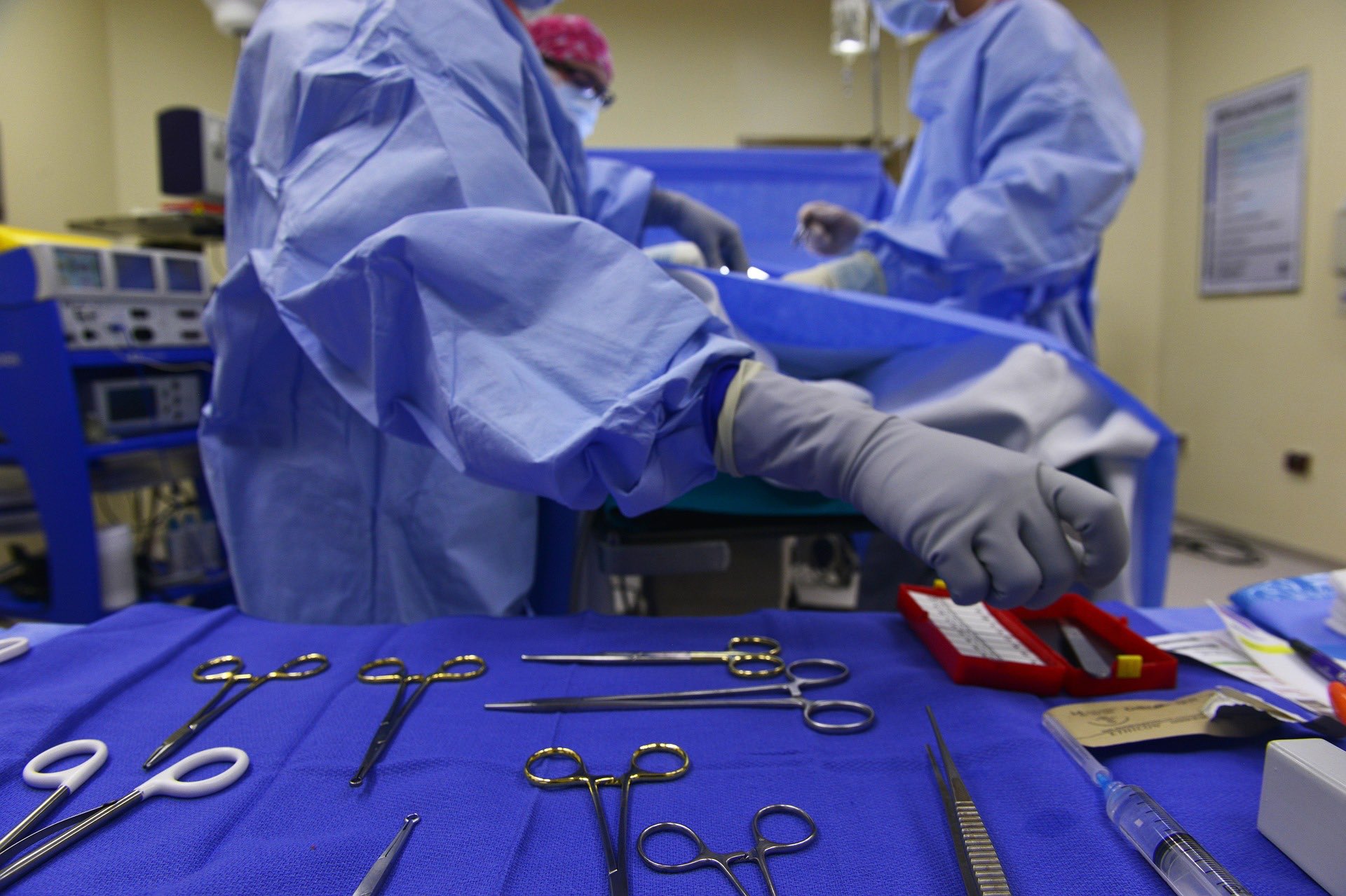
- Guide a needle for biopsy or tumor treatment
- Examine a breast lump
- Check the thyroid gland
- Find genital and prostate problems
- Assess joint inflammation (synovitis)
- Evaluate metabolic bone disease
Risks of Ultrasound
Diagnostic ultrasound is a safe procedure that uses low-power sound waves. There are no known risks.
What are the limitations of Ultrasound?
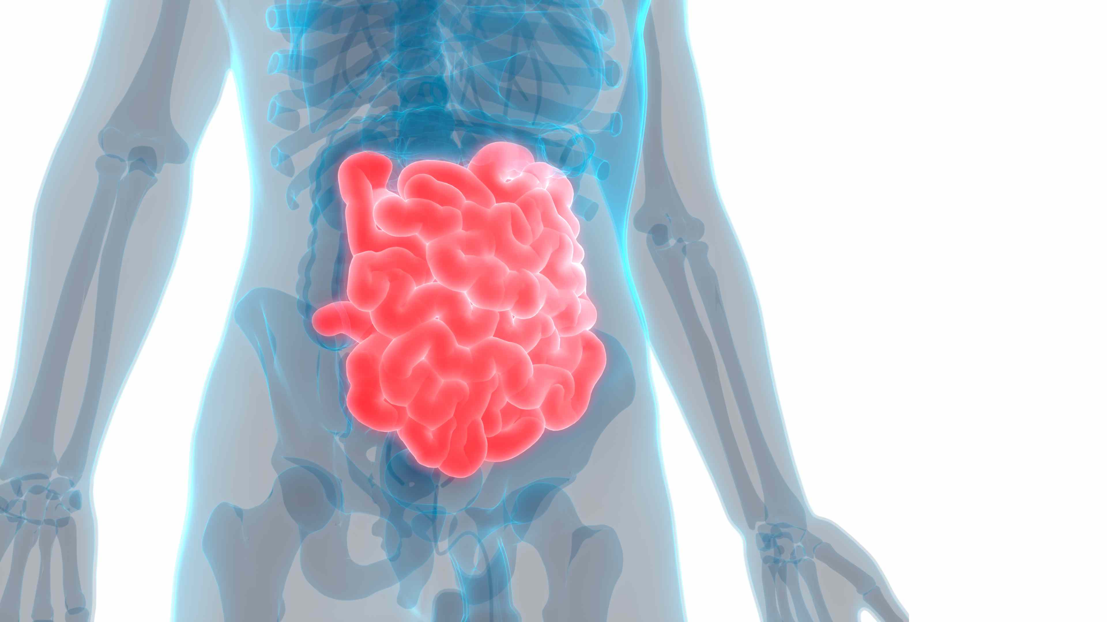
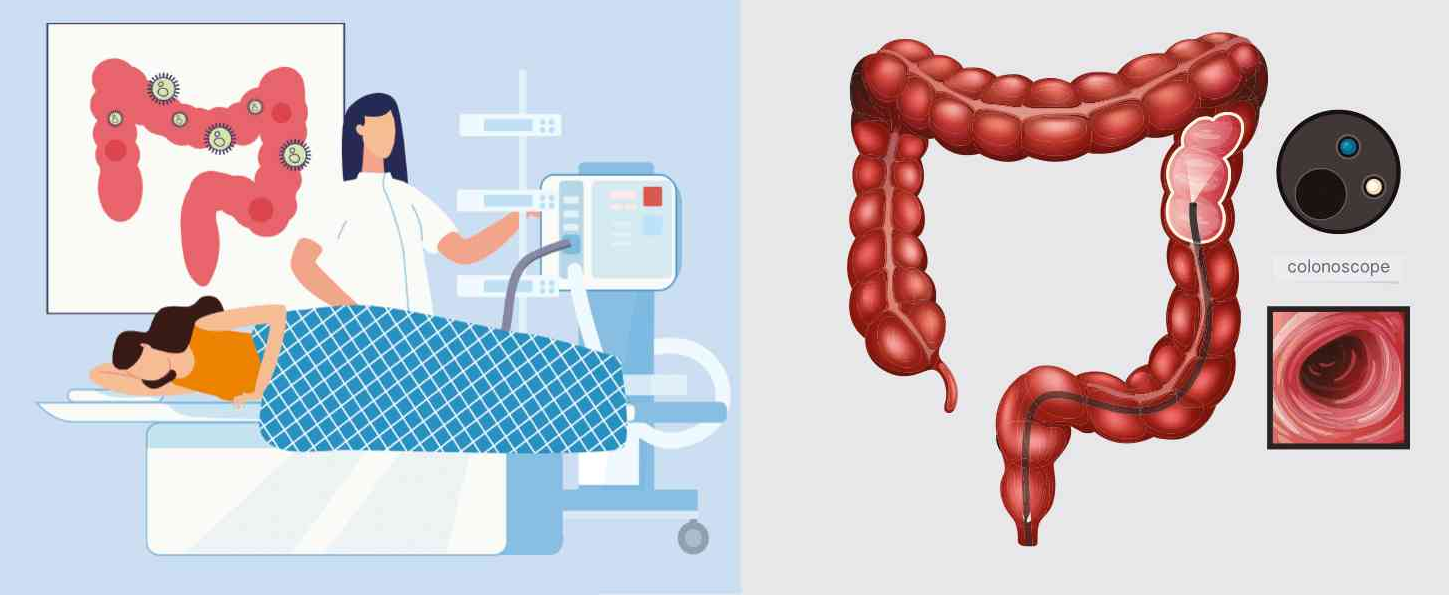
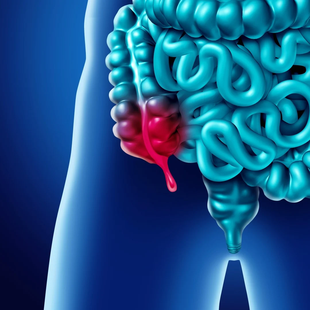
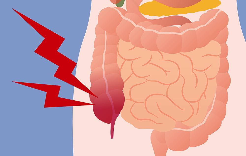
Ultrasound waves are disrupted by air or gas. Therefore, ultrasound is not an ideal imaging technique for the air-filled bowel or organs obscured by the bowel. Ultrasound is not as useful for imaging air-filled lungs, but it may be used to detect fluid around or within the lungs. Similarly, ultrasound cannot penetrate bone, but may be used for imaging bone fractures or for infection surrounding a bone.
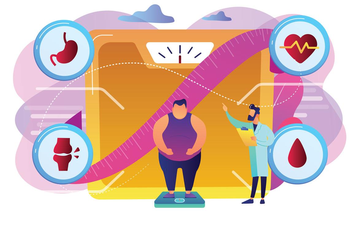
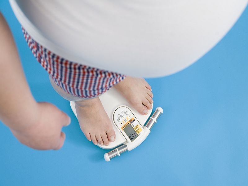
Large patients are more difficult to image by ultrasound because greater amounts of tissue weaken the sound waves as they pass deeper into the body and need to return to the transducer for analysis.
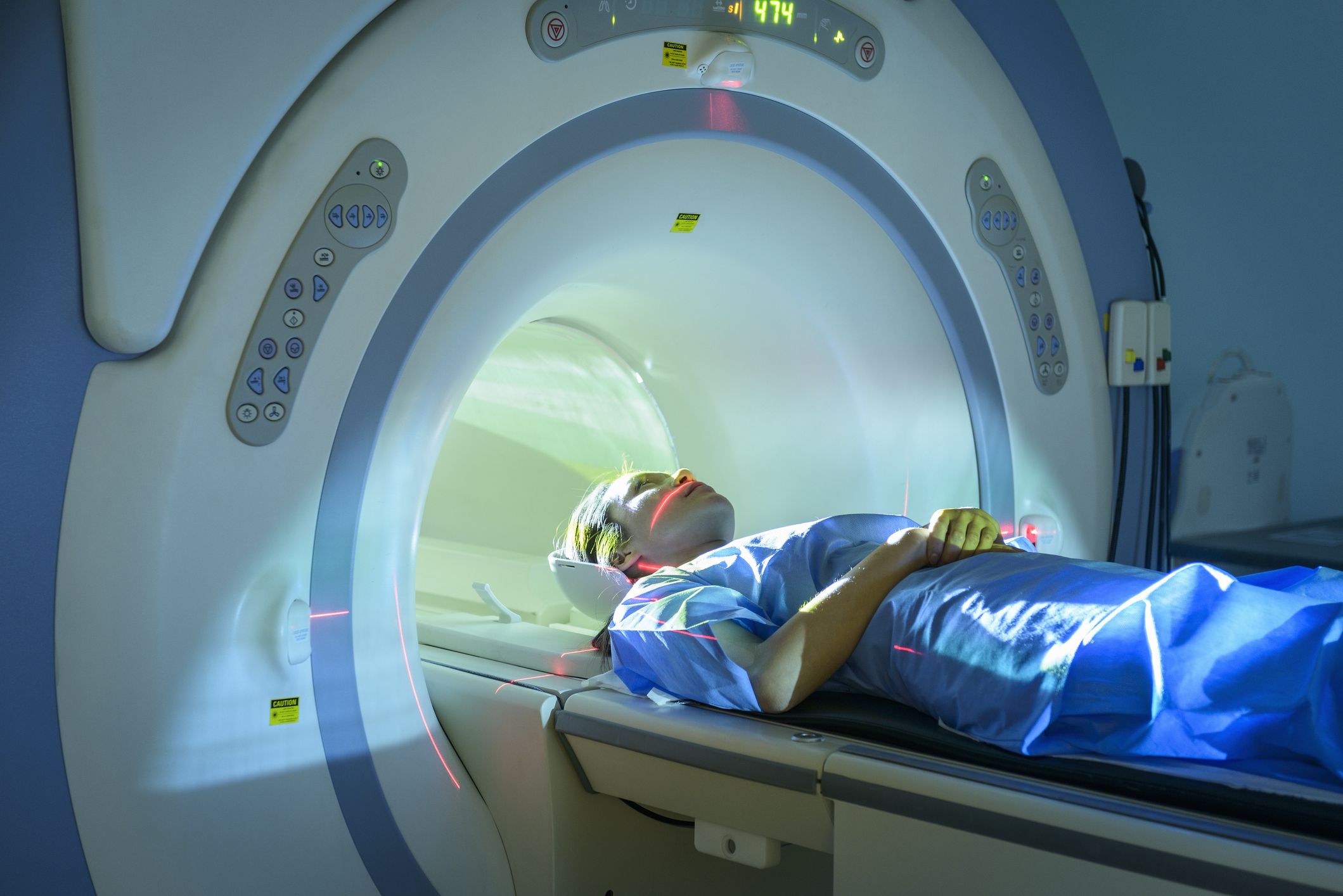
Ultrasound has difficulty penetrating bone and, therefore, can only see the outer surface of bony structures and not what lies within (except in infants who have more cartilage in their skeletons than older children or adults). Doctors typically use other imaging modalities such as MRI to visualize the internal structure of bones or certain joints.
How to prepare for an Ultrasound
Most ultrasound exams require no preparation. However, there are a few exceptions:
- For some scans, such as a gallbladder ultrasound, your care provider may ask that you not eat or drink for a certain period of time before the exam.
- Others, such as a pelvic ultrasound, may require a full bladder. Your doctor will let you know how much water you need to drink before the exam. Do not urinate until the exam is done.
- Young children may need additional preparation. When scheduling an ultrasound for yourself or your child, ask your doctor if there are any specific instructions you'll need to follow.
Clothing and personal items
Wear loose clothing to your ultrasound appointment. You may be asked to remove jewelry during your ultrasound, so it's a good idea to leave any valuables at home.
What to expect when undergoing Ultrasound
Before the procedure
Before your ultrasound begins, you may be asked to do the following:
- Remove any jewelry from the area being examined.
- Remove or reposition some or all of your clothing.
- Change into a gown.
You'll be asked to lie on an examination table.
During the procedure
Gel is applied to your skin over the area being examined. It helps prevent air pockets, which can block the sound waves that create the images. This safe, water-based gel is easy to remove from skin and, if needed, clothing.
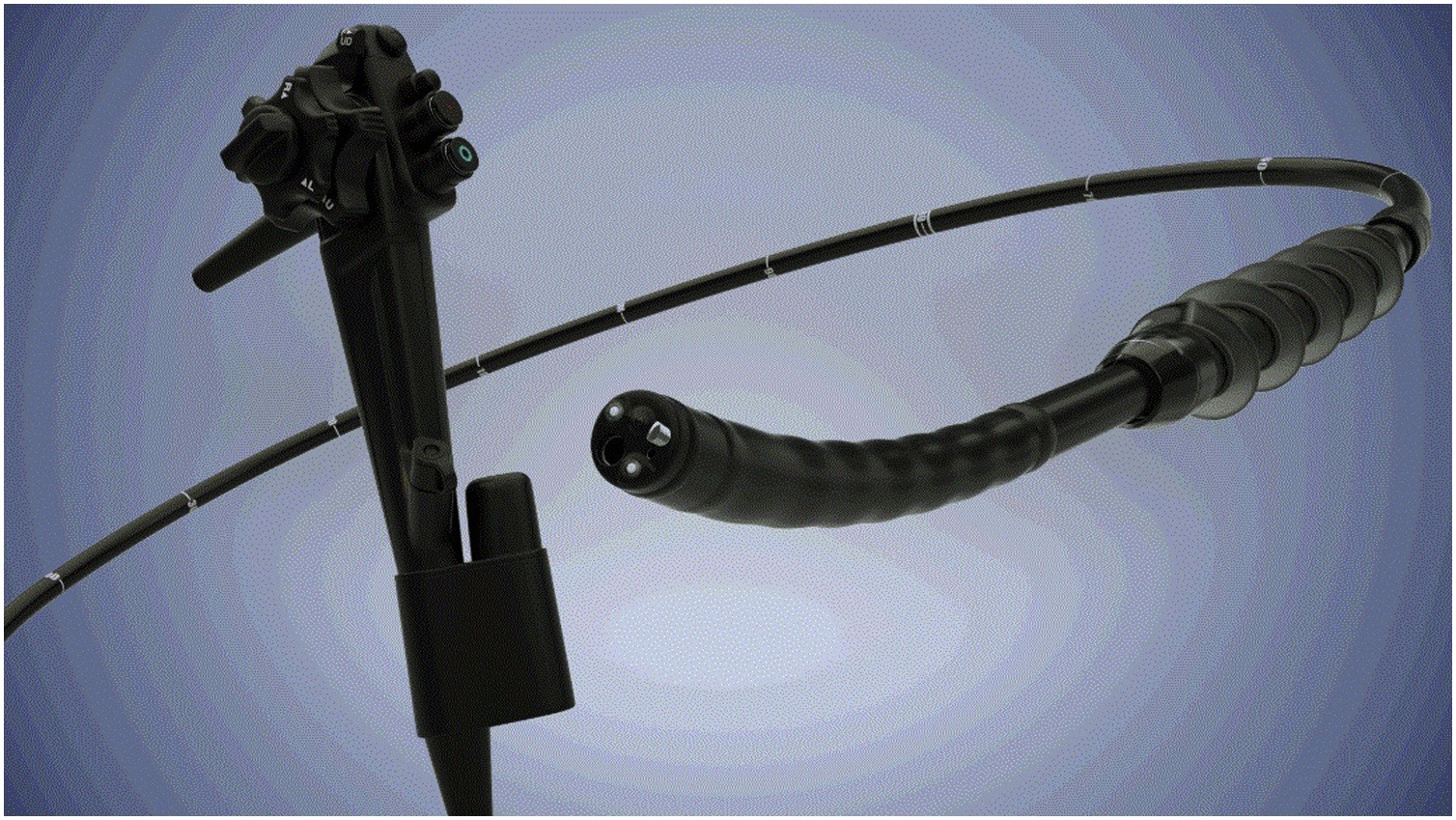
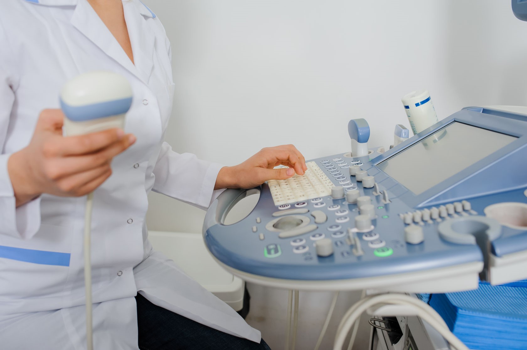
A trained technician (sonographer) presses a small, hand-held device (transducer) against the area being studied and moves it as needed to capture the images. The transducer sends sound waves into your body, collects the ones that bounce back and sends them to a computer, which creates the images.
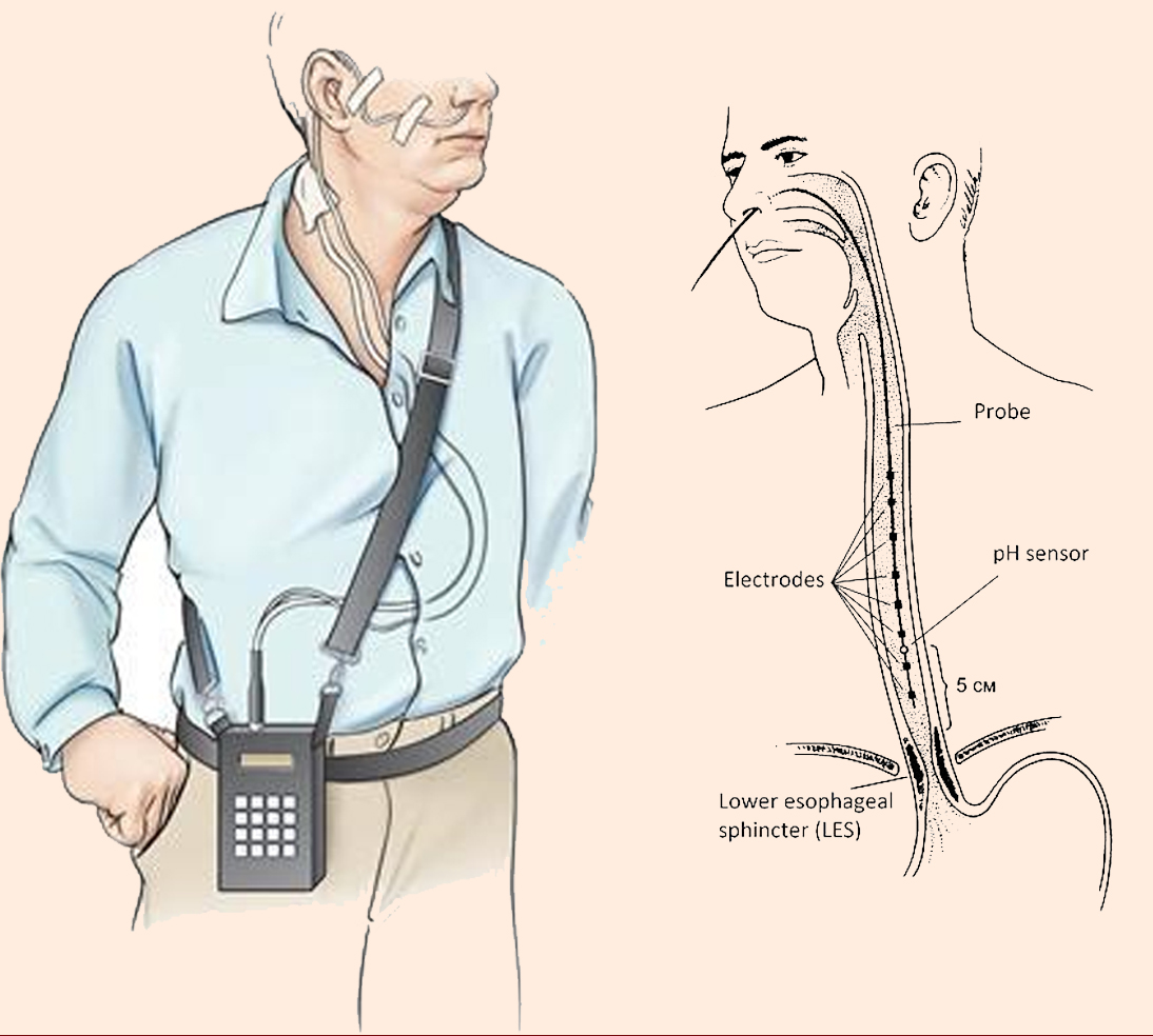
Sometimes, ultrasounds are done inside your body. In this case, the transducer is attached to a probe that's inserted into a natural opening in your body. Examples include:
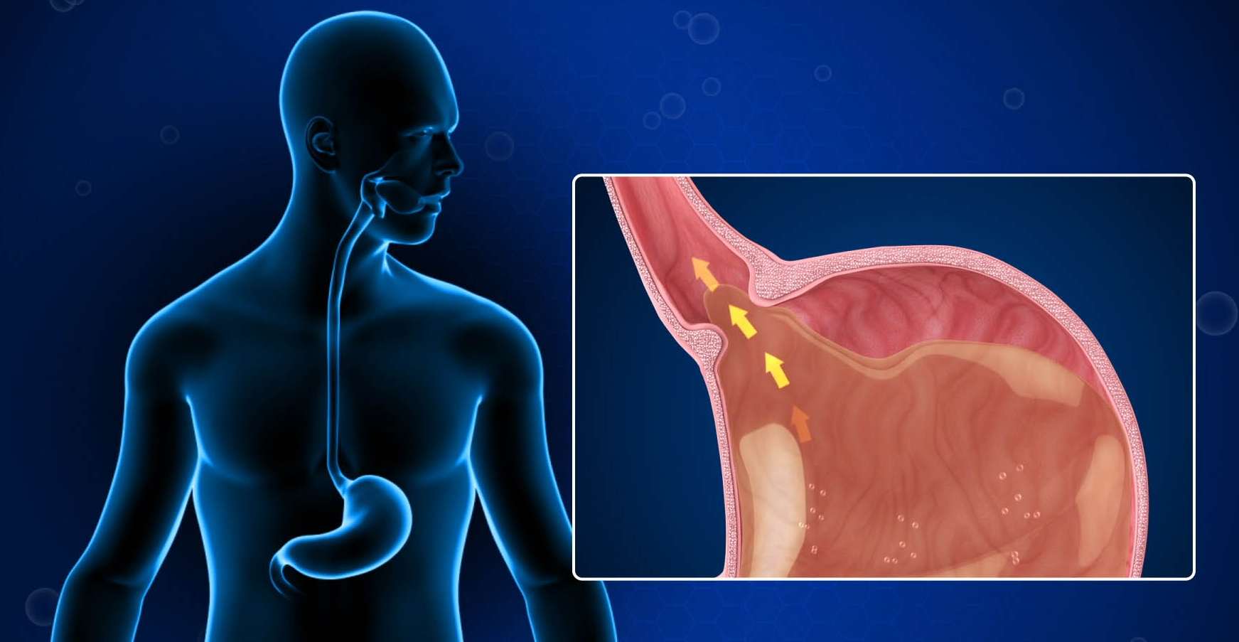
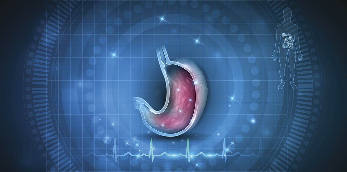
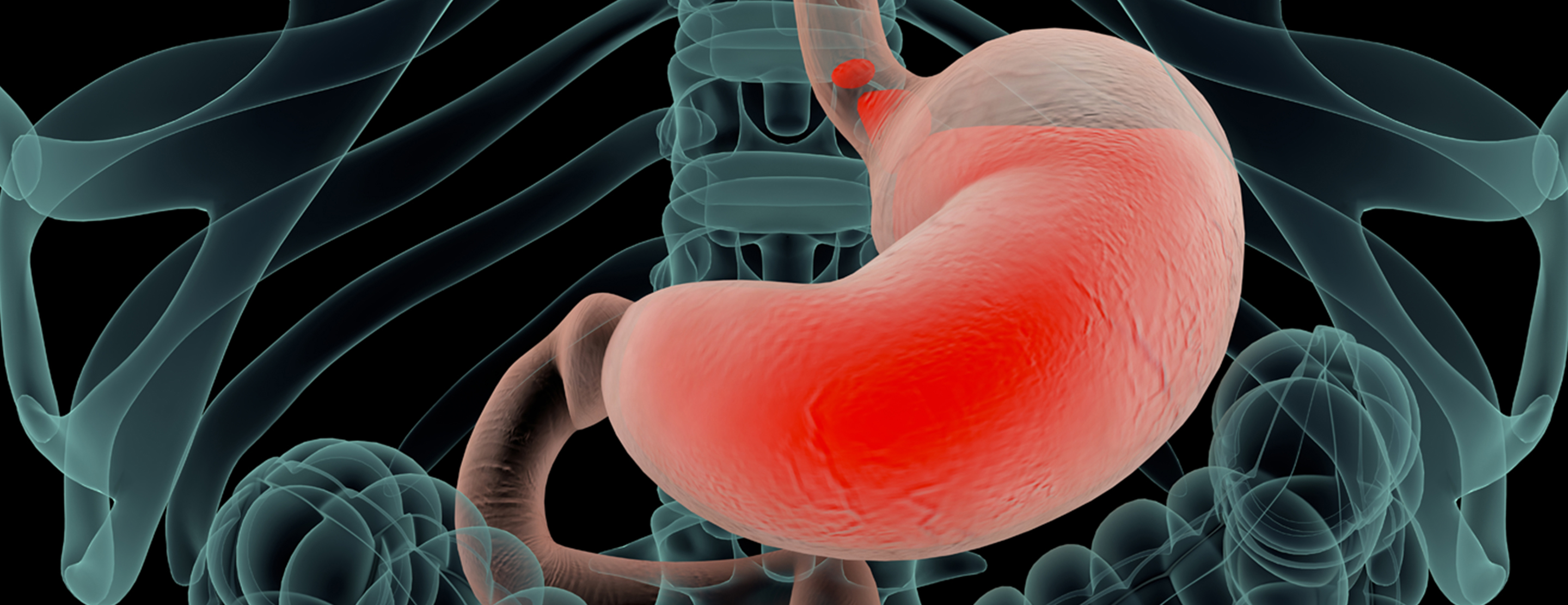
- Transesophageal echocardiogram. A transducer, inserted into the esophagus, obtains heart images. It's usually done while under sedation.
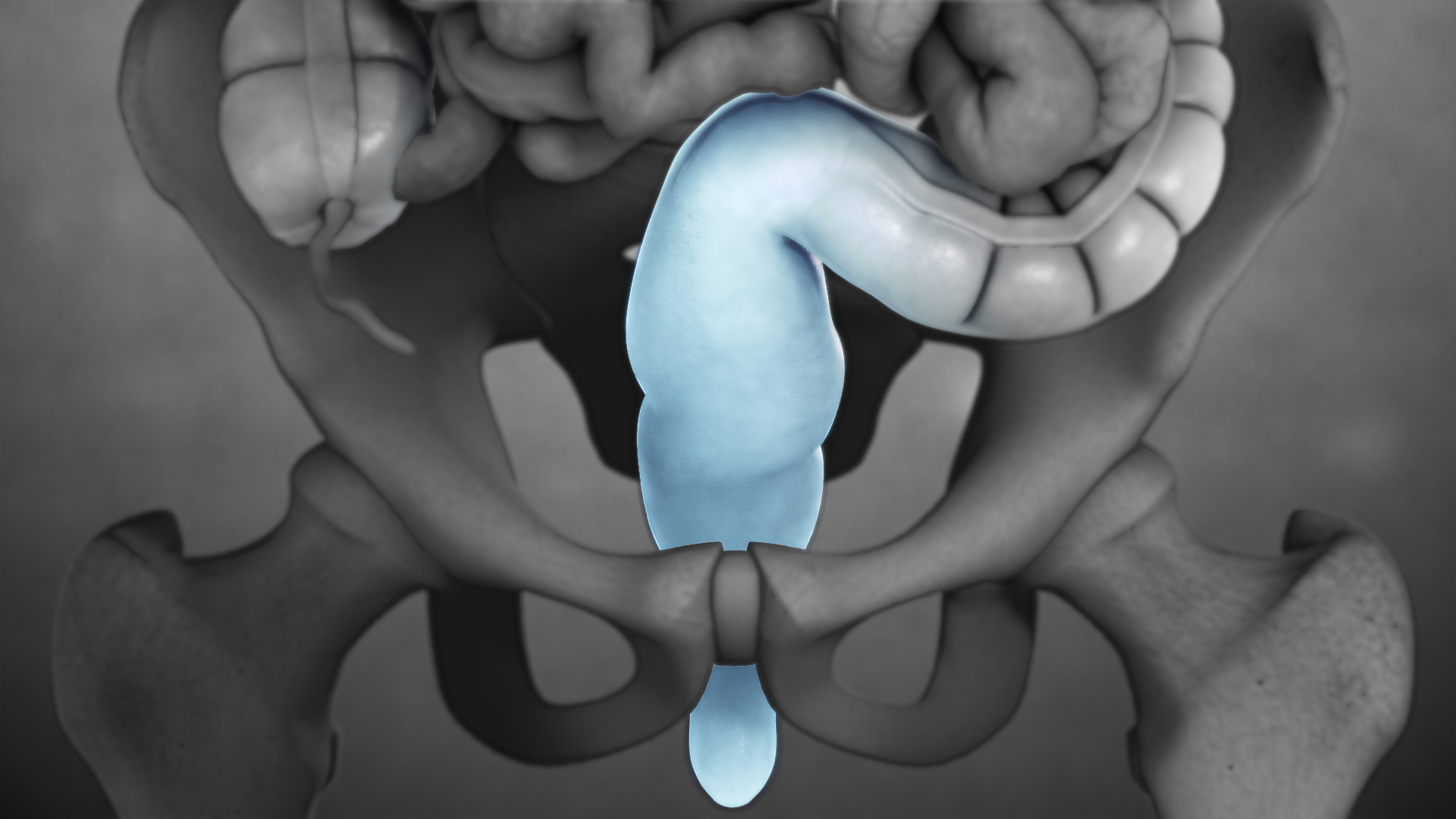
- Transrectal ultrasound. This test creates images of the prostate by placing a special transducer into the rectum.
- Transvaginal ultrasound. A special transducer is gently inserted into the vagina to look at the uterus and ovaries.
Ultrasound is usually painless. However, you may experience mild discomfort as the sonographer guides the transducer over your body, especially if you're required to have a full bladder, or inserts it into your body.
A typical ultrasound exam takes from 30 minutes to an hour.
Results
When your exam is complete, a doctor trained to interpret imaging studies (radiologist) analyzes the images and sends a report to your doctor. Your doctor will share the results with you.
You should be able to return to normal activities immediately after an ultrasound.